Computed tomography (CT) and Magnetic resonance imaging (MRI) each have their own advantages:
Image Acquisition: A CT scanner uses x-rays, a type of ionizing radiation, to acquire images. MRI uses a strong magnetic field and non-ionizing radio frequency (RF) signals to acquire its images. Thus, CT is a good tool for examining tissue composed of elements of a relatively higher atomic number (bone and calcified disks=calcium based) than the tissue surrounding them (flesh=carbon based). MRI is better for viewing non-calcified tissue such as muscle, tendons, soft herniated disks and edema.
Plane of View: CT images are primarily viewed in one plane, transverse. MR images can be viewed in any plane without moving the patient: transverse, sagittal, dorsal and oblique. These different views may help identify a lesion.
Contrast Media: CT may be enhanced by the use of contrast agents (e.g. iodine, barium) containing elements of a higher atomic number than the surrounding tissue. Contrast agents for MRI are those which have paramagnetic properties (e.g. gadolinium).
Convenience: It may be necessary to perform a myelogram prior to spinal CT imaging in order to view compression to the spinal cord. MRI can detect the subtle differences in tissue contrast without the use of myelography. Therefore, a typical spinal procedure using MRI is less invasive and requires less time. However, if a calcified disk is seen on a radiograph which is compressing the spinal cord, a spinal CT may be performed instead of an MRI.
Scanning Parameters: CT uses x-ray attenuation to generate image contrast while MRI uses magnetic fields to generate image contrast. By variation of scanning parameters, a skilled MRI technician can alter and enhance tissue contrast in various ways to detect different features. The different types of images available and the constant advancements in MRI technology have allowed for a seemingly endless list of specialized MRI scans.
Example of Imaging Differences Between CT and MRI:
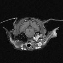 | 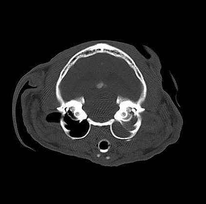 |
| Image 1: Axial T1 MR Image of the Bulla | Image 2: Axial CT Image of the Bulla |
In the case above, “Cheyanne” a 15 year old feline was seen at the Veterinary Neurological Center due to a balance disorder. MRI clearly identified an infection in the right ear canal (Image 1) and there did not appear to be destruction to the bone. If there was destruction to the bone, then the lesion could be invading the brain, causing the prognosis to be very poor. To be certain, a post-MR CT was performed (Image 2) and revealed that the bone was intact. Thus, the neurologist was able to recommend future treatment and possible surgery. This is a good example how CT and MRI are often used in conjunction with one another to reveal a proper diagnosis.
This information is meant to be a guide and not a substitute for veterinary care.
Always follow the instructions provided by your veterinarian.